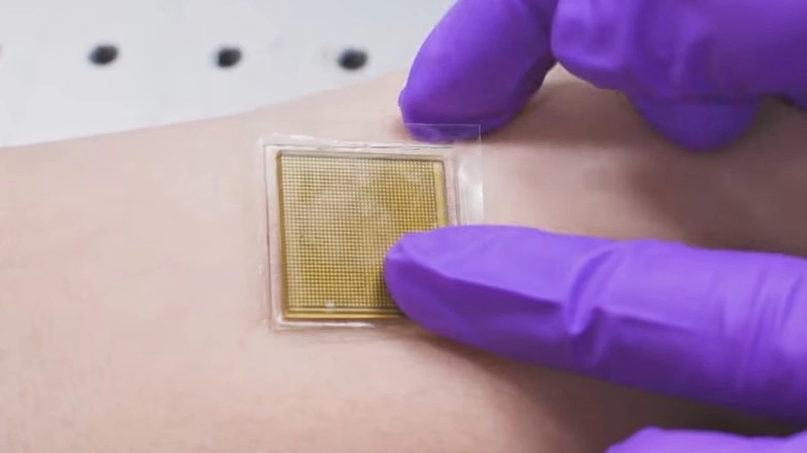
WEB DESK, July 30(ABC): In the field of medical science, an ultrasound is a widely used method of sending sound waves through a body to produce images of internal organs and tissues. It generally requires the use of bulky and expensive machinery as well as expert supervision. A team of engineers at MIT has created a stamp-sized skin patch that will do the demanding job with ease, providing a clear look at internal organs like the lungs and heart in real-time.
The patch is a rigid piezoelectric probe array that attaches to the skin via a transparent gel material. Once attached, it can provide a continuous look at the human body’s vital innards for approximately 48 hours. A doctor or technician can easily apply this patch over the body part that needs scanning.
Once the patch has been applied to the right spot, it is connected to a machine that records the ultrasound signal and converts it into a visible image. Detailed in a paper published in Science, the patch is a remarkable non-invasive technique for imaging body parts and organs that could very well revolutionize the sector.
During the test phase, the ultrasound patch was able to stream a live view of a patient’s abdominal parts, heart, and lungs. The hydrogel elastomer adhesive used in testing was (and is) strong enough that a person can wear the patch while engaged in activities like running, riding a bike, or lifting weights. A key advantage of the ultrasound sticker is that it won’t require continuous supervision by a technician, human or robotic.


